
Ultrasound-Guided Peripheral IV Access in Difficult Cases
Ultrasound significantly enhances venous cannulation in patients with challenging IV access, such as those with obesity, renal failure, or a history of vascular surgeries and graft placements. These patients often present with limited viable access sites, making image-guided techniques particularly valuable.
Anatomical Considerations:
Cephalic Vein: Courses along the lateral aspect of the biceps and continues down the radial side of the forearm. It is typically large, superficial, and often used for PICC line placement.
Basilic Vein: Travels medially along the biceps and forearm, merging with the cephalic vein to form the antecubital “Y.” It terminates on the ulnar side of the hand.
If standard sites are not accessible, scanning the forearm with a systematic approach can help identify alternative veins.
Technique:
Probe Selection: Use a high-frequency linear transducer.
Depth Settings: Begin with a depth of 1–2 cm; adjust for larger patients as needed.
Compression Testing: With a tourniquet applied, gently compress suspected vessels to confirm vein identity and differentiate from tendons or arteries.
Doppler Mode: Utilize color Doppler to confirm venous flow direction and velocity.
Needle Guidance: The long-axis view paired with an in-plane needle approach allows real-time visualization of cannulation.
Equipment Considerations: Arterial catheters with guidewires can be repurposed for deeper veins due to their length and wire-guided access, enhancing procedural success.
Securing Access: If performed under sterile conditions, secure the line with a suture to minimize dislodgement risk.
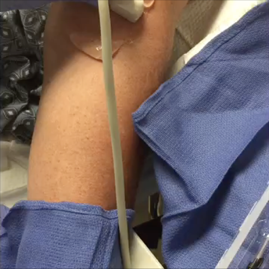
Visual Reference:
The accompanying video demonstrates a long-axis view of the cephalic vein under tourniquet pressure. Note the gradual cephalad flow of blood as it approaches the constriction point.
This image depicts a successfully placed intravenous catheter within the cephalic vein, visualized in short-axis orientation.
This image demonstrates a long-axis view of a properly positioned intravenous catheter with active intraluminal fluid flow. Turbulent flow is visible at the catheter tip, and Doppler imaging may be used to further confirm patency and directional flow.